Some NLM-NCBI services and products are experiencing heavy traffic, which may affect performance and availability. We apologize for the inconvenience and appreciate your patience. For assistance, please contact our Help Desk at info@ncbi.nlm.nih.gov.

. Author manuscript; available in PMC: 2010 Aug 18.
Published in final edited form as: J Comp Neurol. 2008 Sep 10;510(2):158–174. doi: 10.1002/cne.21784

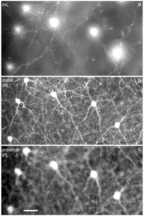
Fig. 2.

Fig. 2

Retinal distribution of DA varicosities. a–c: Confocal images of the mouse retina in three horizontal planes: (a) INL close to the IPL; (b) IPL close to the INL/IPL border; (c) proximal IPL close to the layer of ganglion cells. The six DA perikarya are seen in focus in (b) and out of focus in (a) and (c). Note the presence of DA varicosities at all three levels of the retina. DA, dopaminergic.

HHS Vulnerability Disclosure