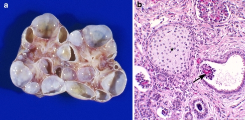
Fig. 3.
An example of glomerulocystic disease in a child with a multicystic dysplastic kidney. a Coronal section of a multicystic dysplastic kidney, b histology demonstrating glomerular cysts (arrow) and a focus of cartilage (asterisk) associated with the multicystic dysplastic kidney (magnification 20×)

