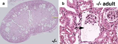
Fig. 6.
Hematoxylin and eosin staining showing glomerulocystic disease in an 8-week old Wwtr1−/− mouse. a Coronal kidney sections showing numerous cysts in the corticomedullary region of the kidney from the knockout mouse (magnification 20×), b higher magnification showing clear glomerulocystic disease with a reasonably normal glomerulus (arrow, magnification 40×). Modified with permission from [36]

