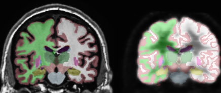
Figure 1a:
(a) Automated, subject-specific segmentation in a 71-year-old male HC subject. Left: MR imaging segmentation. Right: Segmentation applied to coregistered FDG PET volume. ROIs include hippocampus (gold), thalamus (dark green), caudate (blue), putamen (pink), and lateral ventricle (purple). The right and left hemisphere white matter (green and white, respectively) and gray matter (maroon) are shown. (b) Subject-specific cortical parcellation in same subject. Left: Lateral view. ROIs on the lateral surface include inferior (pink), middle (brown), and superior (light blue) temporal cortices; caudal (brown) and rostral (purple) midfrontal cortices; and inferior parietal cortex (violet). Right: Medial view. ROIs on mesial surface include entorhinal cortex (red), parahippocampal cortex (green), four cingulate areas (shades of purple from posterior, posterior middle, caudal anterior, and rostral anterior), and precuneus (lavender).

