
Figure 1b:
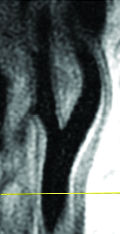
Black-blood MR images through the CCAs of a 71-year-old man. (a, b) Long-axis images through the (a) right and (b) left carotid bifurcation were used to orient subsequent images. (c, d) Contrast material–enhanced T1-weighted transverse images through the (c) right and (d) left CCA were positioned 1.5 cm below the flow divider (yellow lines in a and b). (e, f) Magnifications of c and d. Contours were drawn to delineate the lumen (red circle) and outer wall (green circle) on each image, and the wall was automatically divided into 12 radial segments (white lines). Wall thickness measurements (average of the yellow line thicknesses) and area measurements were generated for each segment and for the entire wall; area measurements were generated for the lumen.