
Figure 2c:
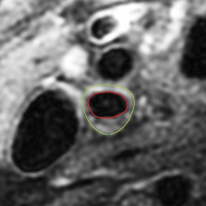
Black-blood MR images obtained through the carotid bifurcation plaque of the same 71-year-old man as in Figure 1. (a) Long-axis image through the thicker carotid bifurcation was selected to orient 16 contrast-enhanced T1-weighted transverse black-blood MR images (yellow lines) centered at the thickest wall or plaque. (b) For this study of remodeling, only the image located one section (2 mm) above the flow divider (blue line in a) was analyzed. (c, d) Contours were drawn to delineate the lumen (red circle), outer wall (green circle), and lipid core (blue line), and the wall was automatically divided into 12 radial segments (white lines in d); thickness and area measurements were generated.