Figure 2.

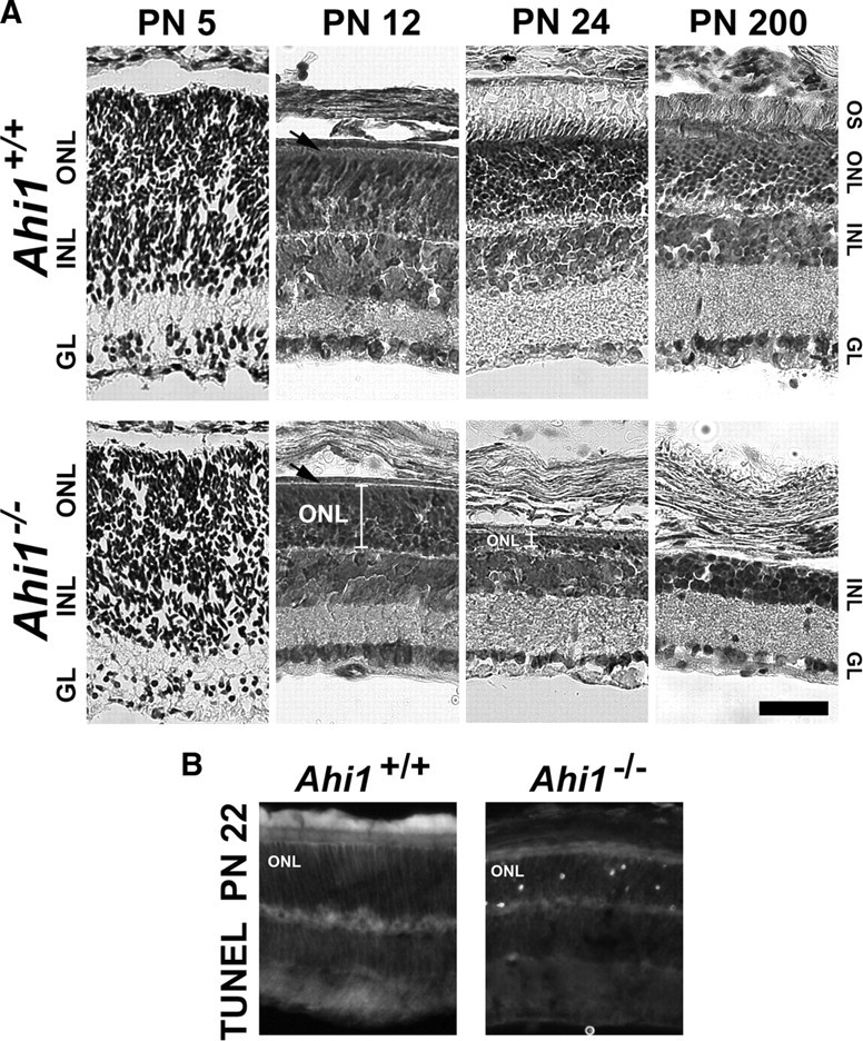
Retinal degeneration in the photoreceptors (outer nuclear layer) of mice with a targeted deletion of Ahi1. A, Cresyl violet staining of retinas from age-matched, wild-type mice (Ahi1+/+; top row) and mice with a targeted deletion of Ahi1 (Ahi1−/−; bottom row) at PN5, PN12, PN24, and PN200, demonstrating the progressive degeneration of the photoreceptor layer in Ahi1−/− retina, but not in Ahi1+/+ retina. The white brackets in the images from the Ahi1−/− retinas at PN12 and PN24 highlight the decrease in size of the photoreceptor layer. The black arrows are pointing to the outer segment layer in Ahi1+/+ retina and the absence of this layer in Ahi1−/− retina. Scale bar, 50 μm. B, TUNEL staining of apoptotic photoreceptor nuclei at PN22 in Ahi1−/− retinas (right), but not in Ahi1+/+ retinas (left). INL, Inner nuclear layer; IGL, ganglion cell layer; ONL, outer nuclear layer; OS, outer segments.
