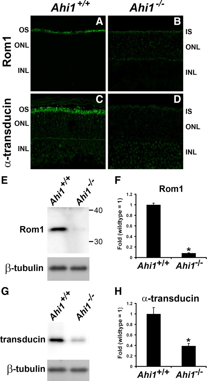
Figure 7.

Decreased levels of photoreceptor outer segment proteins in retinas from Ahi1−/− mice. A–D, Frozen sections of retinas from Ahi1+/+ and Ahi1−/− mice were labeled with antibodies to other photoreceptor outer segment proteins: Rom1 (A, B), and α-transducin (C, D). The outer segment protein Rom1 was mistargeted to the inner segments and cell bodies of photoreceptor cells in Ahi1−/− retinas (B) compared to their outer segment location in the Ahi1+/+ control retinas (A). Also, the levels of Rom1 and α-transducin were significantly reduced in Ahi1−/− retinas (A vs B, C vs D, respectively). E–H, These reductions in Rom1 and α-transducin levels in the photoreceptors were confirmed by Western blotting. The protein levels of endogenous Rom1 and α-transducin from dissected retinas from Ahi1+/+ and Ahi1−/− mice were analyzed by Western blotting (representative blots, E, G) and graphically displayed (F, H; n ≥ 3/genotype). The level of β-tubulin represents the loading control. The error bars represent SEM. Asterisks denote significance from Ahi1+/+ (Rom1, t6 = 23.55, p < 0.0001; α-transducin, t4 = 4.78, p < 0.001). INL, Inner nuclear layer; IS, inner segments; ONL, outer nuclear layer; OS, outer segments.
