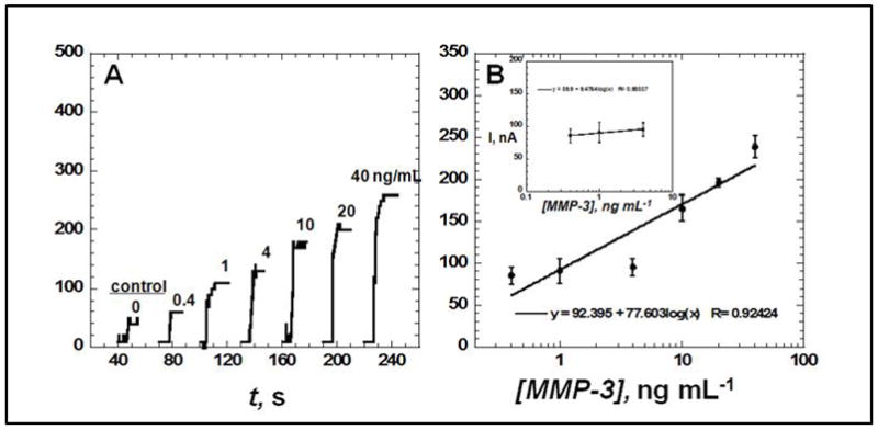
Fig. 2.

Amperometric response for SWCNT immunosensors incubated with MMP-3 (concentration in ng mL−1 labeled on curves) in 10 μL of undiluted newborn calf serum for 1.25 hours, then (A) followed by 10 μL of 200 ng mL−1 biotin–Ab2 in 0.1% Tween-20 for 1.25 h, then 10 μL of streptavidin modified HRP for 30 min, showing current at −0.3 V and 2000 rpm after placing electrodes in buffer containing 1 mM hydroquinone mediator, then injecting H2O2 to 0.4 mM to develop the signal. Controls shown on right with MMP-3 concentrations: full SWCNT immunosensor with 0 ng mL−1 MMP-3 and (B) influence of log MMP-3 concentration on steady state amperometric current for SWNT immunosensor using anti-MMP-3–HRP(14–16). Error bars in part B represent device-to-device standard deviations (n = 3).
