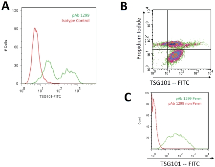
Figure 1.
Flow cytometry analysis of TSG101 antibody binding to the surface of HIV infected cells. Panel A shows MT-4 cells infected with HIV-1NL4-3. Infected (green) and uninfected (red) cells were incubated with polyclonal antibody 1299 (pAb 1299), which is specific to the N-terminal region of TSG101. Membrane integrity of TGS101 positive cells was assessed using propidium iodide as shown in Panel B. The quadrant plot shows MT-4 cells 4 days post infection with HIV-1NL4-3. The lower right quadrant shows cells cell surface expression of TSG101 on cells that exclude PI. The upper right quadrant shows cells with compromised membrane integrity as evidenced by the uptake of PI. Panel C shows lack of binding of 1299 to uninfected MT-4 cells (Red) in the absence of permeabilization reagent, and positive binding when the cells were permeabilized (Green).

