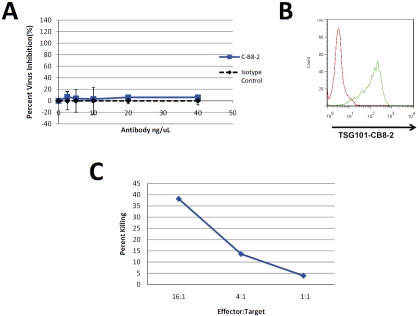
Figure 7.
Antiviral Mechanism of CB8-2 Involves ADCC. Panel A MT-4 cells were infected HIV-1NL4-3 and incubated for 5 days in the presence or absence of C-B8-2 or an isotype control antibody. Supernatants were collected from the MT-4 cultures and virus production was determined by luciferase expression of TZM-bl cells exposed to the culture supernatants. Panel B. Flow cytometry analysis on the infected MT-4 cells used in the virus inhibition experiments in Panel A to demonstrate the level of TSG101 cell surface exposure. Panel C shows the results of a cytoxicity assay exposing HIV-1 infected MT-4 cells to C-B8-2 (5 ng/mL) antibody and varying amounts of NK cells. The levels of cyto-toxicity were determined by measuring LDH release as a result of cell death. Results are represented as percent killing as compared to a total cell lysis control to which lysis buffer was added.

