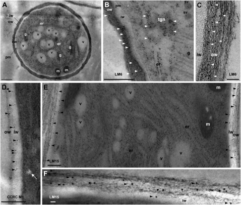
Figure 2.
Electron micrographs showing the ultrastructure (A) and immunogold labeling of cell wall epitopes (B–F) of high-pressure frozen/freeze-substituted Arabidopsis pollen tube grown in vitro for 6 h. A, Cross section of a pollen tube showing the cell wall (cw) composed of two distinct layers: a fibrillar outer wall (ow) and a weakly electron-dense inner wall (iw). Well-preserved organelles are also clearly distinguishable, including endoplasmic reticulum (er), Golgi stacks (g), mitochondria (m), and vacuoles (v). pm, Plasma membrane. B and C, Immunogold labeling of (1→5)-α-l-arabinan epitopes with LM6. Gold particles (arrowheads) are mostly localized in the outer wall layer. In B, possible trans-Golgi network (tgn) and secretory vesicles (sv) labeled with LM6 are seen. D, Immunogold labeling of fucosylated XyG motif recognized by CCRC-M1 in the inner and outer wall layers. Note the presence of gold particles in vesicles in the vicinity of the plasma membrane (white arrow). E and F, Immunogold labeling of nonfucosylated XyG motif (XXXG) with LM15. Gold particles (arrowheads) are visible in the inner and mainly in the outer walls. Bars = 1 μm (A), 0.5 μm (B, D, and E), and 100 nm (C and F).

