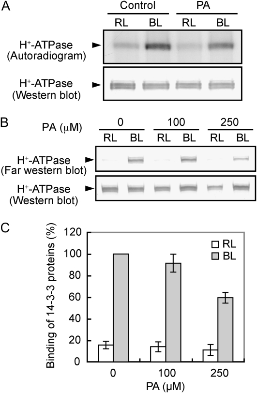
Figure 4.
Inhibition of blue light-dependent phosphorylation of the plasma membrane H+-ATPase and the binding of a 14-3-3 protein to the H+-ATPase by PA. A, Levels of phosphorylation (top) and the amount of the H+-ATPase (bottom) in the presence or absence (Control) of PA. Guard cell protoplasts were preincubated with [32P]orthophosphate for 90 min under red light (RL; 600 μmol m–2 s–1) and then illuminated with a pulse of blue light (BL; 100 μmol m–2 s–1, 30 s). PA at 250 μm was added 30 min before the application of blue light. Reactions were terminated 2.5 min after the blue light. Autoradiography was carried out against H+-ATPase, which was obtained by immunoprecipitation from 100 μg of guard cell proteins. Experiments repeated on three occasions gave similar results. B, Levels of 14-3-3 protein binding to the H+-ATPase in response to blue light. Guard cell protoplasts were treated as described for Figure 3, and the reactions were terminated 2.5 min after the blue light pulse. The binding of a 14-3-3 protein to the H+-ATPase was determined by far western-blot analysis using a recombinant glutathione S-transferase-14-3-3 protein as a probe. C, Quantification of the relative binding of 14-3-3 proteins to the H+-ATPase using ImageJ software. Values shown are means ± se (n = 3).

