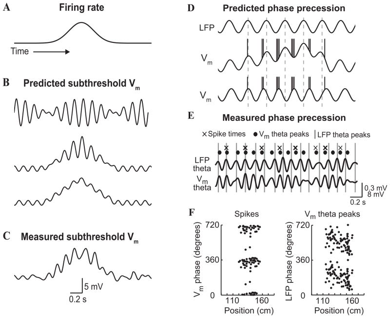
Figure 16. Phase coding by place cells in the hippocampus of mouse performing a virtual navigation task.
(A) Schematic of a place cells firing rate. (B) Schematics of predicted subthreshold membrane potentials (aligned to A) from three different models. Top: in a dual oscillator interference model, two sets of theta-modulated inputs at different frequencies interfere to create a beat-like pattern of membrane potential fluctuations. Middle: in a soma-dendritic interference model, the cell receives theta-modulated inhibitory and excitatory inputs. In the place field, the excitatory drive increases, resulting in a ramp-like depolarization and an increase in the amplitude of excitatory theta oscillations. Bottom: in a network model, a ramp of excitatory drive interacts with theta-modulated inhibitory inputs. (C) Example of a subthreshold membrane potential (filtered from DC-10 Hz) recorded intracellularly from a place cell. Note the simultaneous ramp of depolarization and increase in theta oscillation amplitude. Scale bars refer to the experimentally measured trace only. (D) Schematics of the LFP theta rhythm (Top) and predicted relationships between intracellular and LFP theta to account for phase precession (Middle and Bottom). Middle panel: intracellular and LFP theta have the same frequency. Phase precession of spikes occurs relative to both intracellular and LFP theta owing to a ramp of depolarization. An asymmetric ramp is shown. Bottom panel: intracellular theta in the place field has a higher frequency than LFP theta. Spikes precess relative to LFP theta but not intracellular theta. (E) Filtered (610 Hz) membrane potential and LFP traces in the place field from a simultaneous LFP and whole-cell recording. The times of LFP theta peaks (grey lines), intracellular theta peaks (circles) and spikes (crosses) are shown to illustrate the phase precession of spikes and intracellular theta relative to LFP theta oscillations, and the absence of phase precession of spikes relative to intracellular theta oscillations. (F) Phase precession of spike times relative to intracellular theta (left) and phase precession of intracellular theta peak times relative to LFP theta (right) from a simultaneous LFP and whole-cell recording. Adapted with permission from [422].

