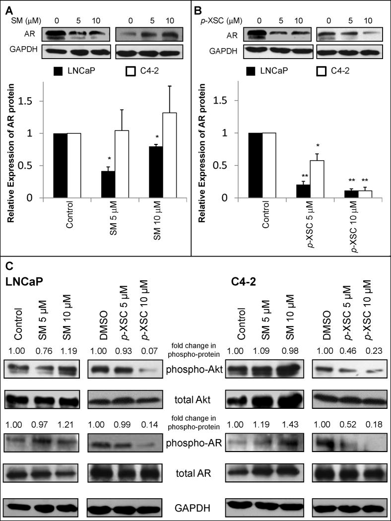
Fig. 3.
The effects of SM and p-XSC on protein expression and phosphorylation. Androgen receptor (AR) protein levels in whole cell lysates from LNCaP and C4-2 cells treated for 24 h with 5 or 10 μM A. SM and B. p-XSC were measured by immunoblot analysis. Results are presented as representative blots from single experiments and in graph form as the average band density (normalized to GAPDH protein levels) from three experiments relative to control samples. (*p<0.05, **p<0.01) C. Levels of phosphorylated Akt and phosphorylated androgen receptor in whole cell lysates of LNCaP and C4-2 cells treated for 1.5 h with 5 or 10 μM SM and p-XSC were measured by immunoblotting. Fold change in band densities of phosphorylated proteins were normalized to the band densities of their respective total protein and to GAPDH levels.

