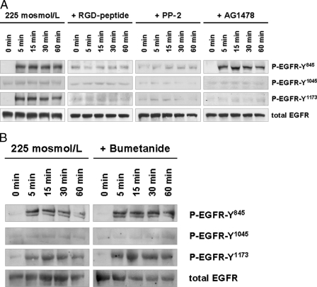
FIGURE 4.
Hypoosmolarity-induced EGFR phosphorylation in perfused rat liver. Rat livers were perfused as described under “Experimental Procedures.” When indicated, RGD peptide (10 μmol/liter), PP-2 (250 nmol/liter), AG1478 (1 μmol/liter) (A) or bumetanide (5 μmol/liter) (B) was added 30 min prior to the institution of hypoosmolarity (225 mosmol/liter). Liver samples were taken at the time points indicated, and phosphorylation of EGFR Tyr845, Tyr1015, and Tyr1173 was analyzed by use of phospho-specific antibodies. Total EGFR served as loading control. Representative Western blots of three independent perfusion experiments are shown. Within 5 min hypoosmolarity induced an RGD peptide- and PP-2-sensitive phosphorylation of EGFR residues Tyr845 and Tyr1173, whereas no phosphorylation at position Tyr1045 became detectable within 60 min (A). AG1478 blunted insulin-induced Tyr1173 phosphorylation, whereas Tyr845 phosphorylation remained unchanged (A), suggestive of an EGFR Tyr845 transphosphorylation leading to EGFR Tyr1173 autophosphorylation as observed upon insulin perfusion (Fig. 1). In contrast to insulin-induced EGFR phosphorylation, bumetanide did not affect hypoosmotic-induced EGFR phosphorylation (B).

