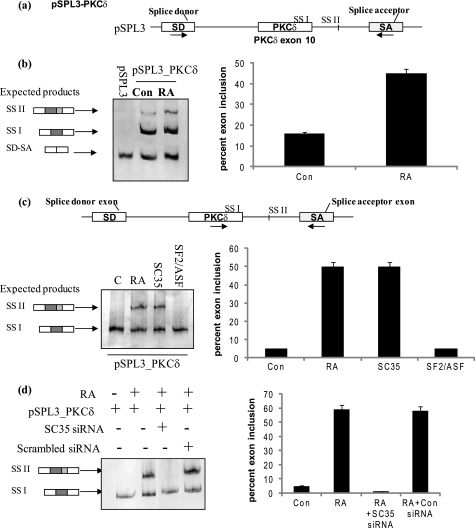
FIGURE 6.
Minigene analysis demonstrates that RA promotes utilization of 5′ splice site II on PKCδ exon 10 pre-mRNA. a, schematic represents PKCδ pre-mRNA exon 10 and flanking introns cloned into pSPL3 splicing vector between the SD and SA exons. The resulting minigene is referred to as pSPL3_PKCδ minigene. Arrows indicate position of primers used in RT-PCR analysis. b, pSPL3_PKCδ minigene and pSPL3 empty vector were transfected overnight, and then the cells were treated with or without 10 μm RA for 24 h. Total RNA was extracted and RT-PCR performed using primers as described above. Expected products are SD-SA: constitutive splicing; SSI: usage of 5′ splice site I; SSII: usage of 5′ splice site II. c, 2 μg of SC35 or SF2/ASF was co-transfected along with the pSPL3_PKCδ splicing minigene. In separate wells, 10 μm RA was added for 24 h. Total RNA was extracted and RT-PCR performed using PKCδ exon 10 and SA primers as shown in the schematic. SSI: usage of 5′ splice site I; SSII: usage of 5′ splice site II. d, SC35 siRNA (100 nm) or scrambled siRNA was co-transfected with pSPL3_PKCδ minigene. 10 μm RA was added to wells as indicated. Total RNA was extracted and RT-PCR performed using PKCδ exon 10 and SA primers as shown above in c. 5% of the products were separated by PAGE and silver stained for visualization. Graphs represent percent exon inclusion calculated as SS II/(SS II + SSI) × 100 in the samples and are representative of four experiments performed separately. These results demonstrate that co-transfection of SC35 with the pSPL3_PKCδ minigene promotes utilization of 5′ splice site II. Further, RA is unable to promote utilization of 5′ splice site II on PKCδVIII pre-mRNA in the absence of SC35.

