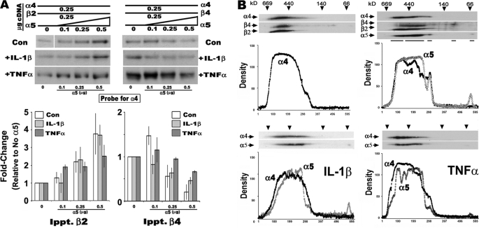
FIGURE 5.
The presence of α5 moderates the influence of the pro-inflammatory cytokines IL-1β and TNFα on nAChR receptor assembly. A, similar to experiments in Fig. 2 only IL-β or TNFα was added for the duration of the transfection (control gels are the same as in Fig. 2). Western blot analysis of cells co-transfected with fixed amounts of α4 and the indicated β subunit and increasing amounts of α5 cDNA. The relative amount of association of α4 with the indicated β subunit is detected using Western blot, and the relative band density is plotted. B, in similar experiments cell membranes were examined by two-dimensional BNG analysis, and the results for just α4 and α5 are shown for clarity. Plots of α4 immunoreactivity in cells treated with either IL-β or TNFα and co-transfected with α5 or not co-transfected (as labeled) are placed below the blots. Control experiments were conducted between three and eight times as in Fig. 2. Cytokine experiments are based upon an n value of 3.

