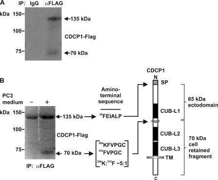
FIGURE 3.
Identification of the sites at which CDCP1 is proteolytically processed. A, anti-Flag Western blot analysis of proteins obtained from mouse IgG and anti-Flag antibody immunoprecipitations from lysates of HeLa-CDCP1-Flag cells treated with PC3 cell-conditioned medium. B, Coomassie-stained PVDF membrane of anti-Flag immunoprecipitates obtained from HeLa-CDCP1-Flag cells either untreated or treated with PC3 cell-conditioned medium. To the right is shown the N-terminal sequence obtained by Edman degradation sequencing of 70 and 135 kDa CDCP1 and a diagram showing the location of the identified N termini within the CDCP1 structure. The data indicated that 135 kDa CDCP1 is processed after Arg-368 and Lys-369 in the ratio 5:1 in a region located between CUB-like domains 1 and 2 (CUB-L1 and CUB-L2). SP, signal peptide; TM, transmembrane domain.

