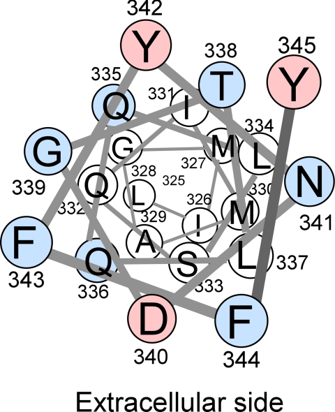
FIGURE 6.
Helical wheel representation of TM7 of Hxt7 shown from the extracellular side. Red circles indicate residues whose single-Cys mutants were inhibited by pCMBS, whereas blue circles represent residues for which Cys substitution resulted in mutants with low activities (<15% of that of Cys-less Hxt7). With the exception of Leu337, all pCMBS-accessible residues and residues sensitive to Cys replacement are located in the outer half of TM7.

