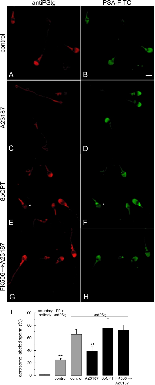
FIGURE 5.
Synaptotagmin dephosphorylation depends on calcium and calcineurin. Nonpermeabilized sperm were incubated for 15 min at 37 °C with 100 μm 2-APB and, when indicated, 1 μm FK 506. The cells were then further incubated for 30 min at 37 °C with no additions (control, A and B) or stimulated with 10 μm A23187 (C, D, G, and H) or 50 μm 8pCPT (E and F). The cells were then fixed and double-stained with the antiPStg antibody followed by an anti-rabbit Cy3 (A, C, E, and G) and FITC-coupled P. sativum agglutinin to differentiate between reacted and intact sperm (B, D, F, and H). The asterisk in F shows a reacted sperm with equatorial lectin staining and very faint antiPStg labeling. Bar, 5 μm. In I, at least 200 cells treated as described for A–H were classified as having or lacking distinct acrosomal phosphosynaptotagmin staining. The percentage of immunolabeled sperm in five independent experiments was recorded. As controls, the percentages of labeled cells when the antiPStg antibody was excluded or when it was preblocked with excess phosphorylated peptide are shown (n = 2, supplemental Fig. S3). The data represent the means ± S.E. or the means ± range. The asterisks indicate significant differences from control sperm incubated with unblocked antiPStg antibody (p < 0.01, one-way ANOVA and Dunnett test).

