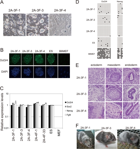
FIGURE 2.
Characterization of the genome integration-free 2A-3FiPS clones. A, morphology of three genome integration-free 2A-3FiPS colonies. B, immunocytochemical detection of Oct3/4 using the h-134 antibody (Santa Cruz Biotechnology). 4′,6-Diamidino-2-phenylindole (DAPI) was used as a nuclear counter stain. C, RT-PCR analysis of ES-marker genes. Error bars indicate standard deviations (n = 3). D, methylation analysis of the Oct3/4 and Nanog gene promoter regions using bisulfite genomic sequencing. Open and closed circles indicate unmethylated and methylated CpGs, respectively. E, teratomas derived from the 3 2A-3FiPS clones. F, a chimeric mouse generated from 2A-3FiPS-1, -3, and -4.

