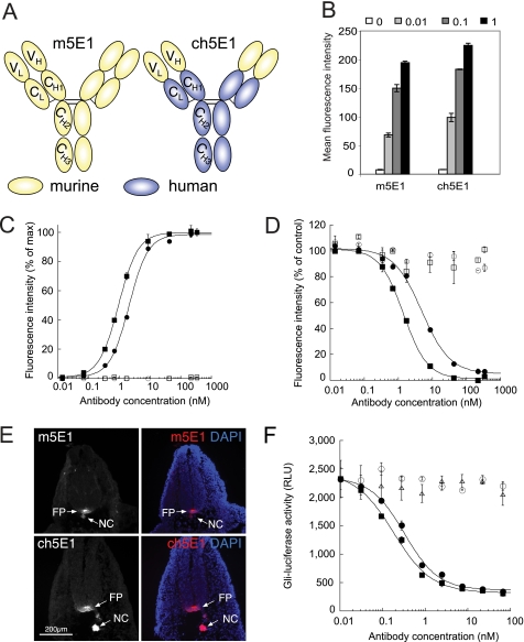
FIGURE 1.
Chimeric 5E1 is functionally equivalent to murine 5E1. A, schematic of murine 5E1 (m5E1, yellow) IgG (33) and its chimeric counterpart (ch5E1), where the constant domains (CH1–3 and CL) have been replaced with the corresponding domains from the humanized antibody trastuzumab (blue) (35), leaving the variable light and heavy (VL and VH) domains of the murine 5E1 antibody intact. B, ch5E1 and m5E1 bind similarly to an endogenous Hh-expressing cell line. Flow cytometry analysis is shown of endogenous Hh in HT29 cells with the indicated concentrations of m5E1 or ch5E1 (in μg/ml). The means ± S.D. of triplicate reactions are plotted. C, ch5E1 (■) and m5E1 (●) bind similarly to stably transfected Shh-COS cells by flow cytometry analysis. The means ± S.D. of a representative duplicate experiment are shown. Isotype controls (chimeric IgG (□) or murine IgG1 (○)) show no appreciable binding. D, ch5E1 and m5E1 compete for cell surface Shh. The ability of increasing amounts of ch5E1 (■) or m5E1 (●) to compete with ∼0.69 nm (0.1 μg/ml) m5E1 for binding to Shh-expressing cells and vice versa as monitored by flow cytometry analysis is shown, normalized to 100% for no competitor after background subtraction. Isotype controls (murine IgG1 (○) or chimeric IgG (□)) are unable to compete for Shh binding. E, ch5E1 specifically detects Hh in the developing mouse embryo. E10.5 embryos were sectioned and stained with m5E1 (top) or ch5E1 (bottom), followed by Cy3-conjugated secondary antibodies (left panel and red in merged right panel) and 4′,6-diamidino-2-phenylindole (DAPI) (blue nuclear staining in right merged panels). ch5E1 is as specific as m5E1 in detecting Hh in the notochord (NC) and floor plate (FP). Scale bar is 200 μm (images taken at ×10 magnification). F, ch5E1 and m5E1 inhibit Hh signaling similarly. HT29 cells secreting Hh were co-cultured with S12 cells (C3H10T1/2 cells stably expressing a Gli-luciferase reporter (36)). Hh signaling was stimulated by serum starvation for 24 h in the presence of the indicated concentrations of ch5E1 (■), m5E1 (●), hIgG1 (▴), or mIgG1 (○) antibodies. The means ± S.D. of the luciferase signals (RLU; relative luminescence units) of triplicate measurements are plotted. This experiment is representative of multiple independent experiments.

