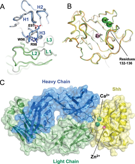
FIGURE 3.
Structure of 5E1 bound to Shh. A, comparison of the structures of ch5E1 Fab alone and in complex with Shh. Shh-bound ch5E1 light and heavy chains are shown as C-α ribbons and colored green and blue, respectively; both heavy and light chains of the free ch5E1 Fab are colored gray. Residues in CDR H3 differing most upon binding to Shh (not pictured) are labeled. B, comparison of the structures of Shh free (white), bound to Cdon (orange), Hhip (green), or 5E1 (yellow). The bound divalent metal cations are shown as spheres (Zn2+ in pink and Ca2+ in green). C, complex between 5E1 and Shh. The Fab is colored as in A, and Shh is in yellow, with Zn2+ and Ca2+ colored as in B.

