FIGURE 5.
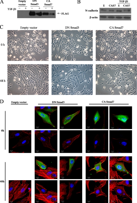
Smad3 mediates TGF-β1-induced actin rearrangement. FLAG-tagged dominant-negative (DN) Smad3 and FLAG-tagged constitutively active (CA) Smad7 constructs were transfected into ARPE-19 cells. Empty vector was used as a control. After transfection, cells were incubated for 48 h with medium alone or medium containing 10 ng/ml TGF-β1, examined by phase-contrast microscopy (C), and then lysed and subjected to Western blot analysis using anti-FLAG antibodies (A). B, Western blot of N-cadherin under the same treatment conditions as A is shown. E, empty vector; CAS7, constitutively active Smad7. D, ARPE-19 cells were transfected for 12 h with plasmids expressing DN Smad3 or CA Smad7. After transfection, cells were switched to serum-free medium for 3 h and treated with TGF-β1 (10 ng/ml) for 48 h. Cells were fixed and stained with anti-FLAG followed by Alexa 488-conjugated secondary antibody (green) and stained with rhodamine-labeled phalloidin (red). Blue is Hoechst nuclear staining. Pictures were taken under a Zeiss confocal microscope. Bar, 10 μm.

