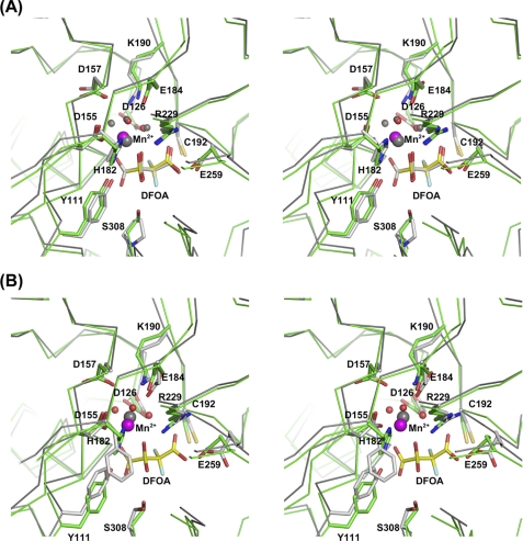
FIGURE 7.
Stereoscopic representations of superposed active sites of OAH and DMML (A) and OAH and PDP (B). The protein backbone Cα atoms are traced, and selected side chains are shown. OAH carbon atoms are colored green, and those of either DMML or PDP are colored gray. A, the DFOA-bound structures of both OAH and DMML are shown. B, the structure of PDP does not include DFOA. Instead, the closed conformation was obtained with glutaraldehyde that reacted with the catalytic cysteine (used to enable flash-cooling of the crystals). For clarity, the glutaraldehyde adduct is not depicted in the image. OAH Mn2+ is colored magenta and that of DMML in gray, as is the Mg2+ in the PDP structure.

