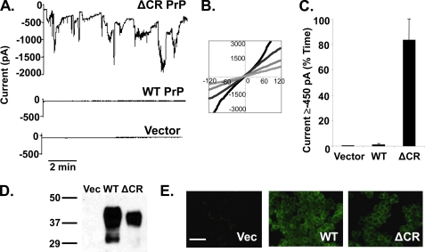
FIGURE 1.
Δ CR PrP induces spontaneous inward currents in HEK cells. A, whole-cell patch clamp recordings were made from HEK cells expressing ΔCR PrP, WT PrP, or vector at a holding potential of −80 mV. B, I-V plots collected at four different times during recording from HEK cells expressing ΔCR PrP. C, current activity recorded from HEK cells expressing vector, WT PrP, or ΔCR PrP was plotted as the percentage of total time the cells exhibited inward current ≥450 pA (mean ± S.E., n = 5 cells) at a holding potential of −80 mV. D, Western blot showing levels of PrP in HEK cells expressing vector (Vec), WT PrP, or ΔCR PrP. Molecular size markers are given in kDa. E, surface immunofluorescence staining of PrP (green) on HEK cells expressing vector, WT PrP, or ΔCR PrP. Scale bar = 50 μm.

