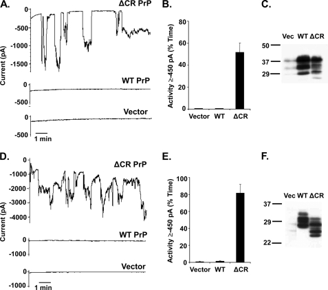
FIGURE 2.
Expression of ΔCR PrP in N2a mouse neuroblastoma cells and Sf9 insect cells produces spontaneous inward currents. A, whole-cell patch clamp recordings at −80 mV of N2a cells 24 h after transfection with plasmids encoding ΔCR PrP, WT PrP, or vector. Transfected cells were recognized by expression of GFP, encoded in a co-transfected plasmid. B, quantitation of the currents shown in panel A, plotted as the percentage of total time the cells exhibited inward current ≥450 pA (mean ± S.E., n = 5 cells). C, Western blot to detect PrP in the N2a cells used in panel A. Vec, vector. D, whole-cell patch clamp recordings at −80 mV of Sf9 cells 36 h after transfection with plasmids encoding ΔCR PrP, WT PrP, or vector. Transfected cells were recognized by expression of GFP, encoded in a co-transfected plasmid. E, quantitation of the currents shown in panel D, plotted as the percentage of total time the cells exhibited inward current ≥450 pA (mean ± S.E., n = 5 cells). F, Western blot to detect PrP in the Sf9 cells used in panel D. PrP expressed in Sf9 cells migrates with a lower Mr than in mammalian cells (HEK or N2a) due to differences between insect and mammalian cells in N-linked glycosylation.

