FIGURE 6.
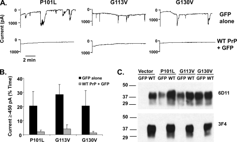
Currents induced by disease-associated point mutants are silenced by overexpression of WT PrP. A, whole-cell patch clamp recordings at a holding potential of −80 mV were made from HEK cells expressing P101L, G113V, or G130V PrP 48 h after infection with lentivirus encoding GFP alone (upper traces) or WT PrP plus GFP (lower traces). B, quantitation of the currents recorded in panel A, plotted as the percentage of total time the cells exhibited inward current ≥450 pA (mean ± S.E., n ≥ 4 cells). Black bars represent GFP alone, and gray bars represent WT PrP plus GFP. C, Western blot showing relative PrP expression levels of each stably transfected cell line after transduction with either WT PrP-encoding or control lentiviruses. Antibody 6D11 recognizes both mutant and WT PrP, whereas antibody 3F4 specifically recognizes WT PrP (which contains the 3F4 epitope).

