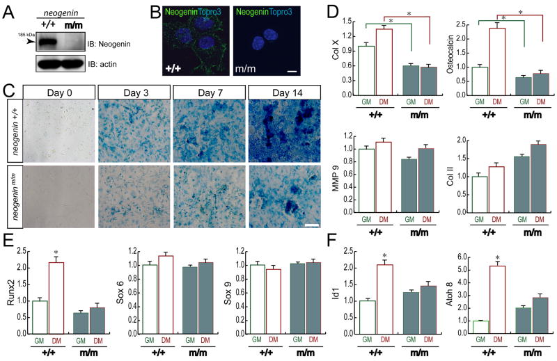
Figure 3. Defective chondrogenesis in vitro in cells from neogenin deficient mice.
Western blot (A) and immunostaining (B) analyses of neogenin expression in wild type (+/+) and mutant (m/m) chondrocytes. Neogenin was detected in wild type, but not mutant, chondrocytes, demonstrating the specificity. Bar, 5 μm. (C) Reduced In vitro chondrocyte differentiation in neogenin deficient cells. Chondrocytes from new born wild type and mutant mice were incubated with differentiation medium (DM, growth medium supplemented with 10 mM β-glyceriophosphate and 50 μg/ml ascorbic acid) for indicated days. Cells were stained with alcian blue to view chondrocyte matrix, a differentiation marker. Bar, 50 μm. (D–F) Real time PCR analysis of genes associated with chondrocyte proliferation and/or differentiation (D), different transcriptional factors known to be important for chondrocyte differentiation (E), and BMP downstream target genes (F) was shown. In (D–F), chondrocytes isolated from new born mice were cultured in the presence of growth medium (GM) or differentiation medium (DM) for 24 hours. RNAs were isolated for real time PCR analysis as described in the Methods. Date were normalized by internal control of GAPDH, and presented as fold over wild type control (mean +/− SD, n = 6); * denoted p<0.05, significant difference from wild type control.

