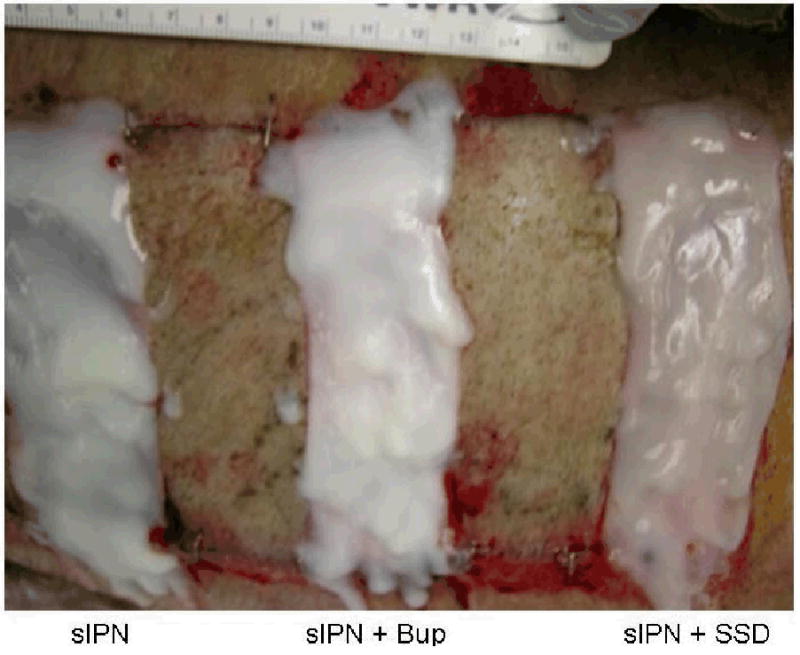
Figure 1.

Example of wound treatment arrangement on pig wounds. Wounds cut at 0.022 in or 0.030 in dermatome cut depth settings were treated with semi-interpenetrating network (sIPN) containing no drug (left), sIPN with 0.5% bupivacaine (Bup) (middle), sIPN with 1% silver sulfadiazine (SSD) (right), and Xeroform™ (not pictured). Alternating split thickness autografts were applied between each treatment to provide physical separation between experimental wound sites.
