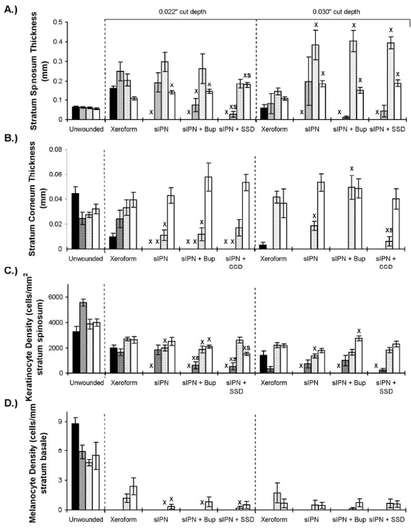
Figure 6.

Epidermal features of wounds dressed with Xeroform™, sIPN, sIPN with 0.5% bupivacaine (Bup), and sIPN with 1% silver sulfadiazine (SSD) examined histologically at 7 (■), 9 ( ), 14 (
 ), and 21 (□) days. The 0.016 in dermatome cut depth wounds are shown on the left, whereas 0.022 in wounds are on the right. Thickness of stratum spinosum(A), thickness of stratum corneum(C), viable keratinocyte density within epidermis(C), and melanocyte density(D) are displayed. Data is reported as average of 10 total viewing regions from biopsies of two separate pigs ± standard error. “X” indicates significant difference from Xeroform™ treated tissue, and “S” indicates significant difference from sIPN without drug treatment (P<0.05).
