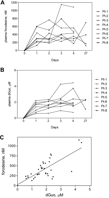
Figure 3.
Plasma forodesine and dGuo levels during therapy. Whole blood was collected from all 8 patients at indicated time points, and plasma was separated. Forodesine (A) and dGuo (B) levels in each sample were determined with the use of a tandem mass spectrometry liquid chromatography as described in “Measurement of plasma dGuo and forodesine.” Each symbol represents a patient. The correlation between plasma forodesine and plasma dGuo (C) was evaluated by plotting data from panels A and B, and linear regression analysis was performed.

