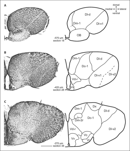
Fig. 1.
Photomicrographs and corresponding line drawings of the A. burtoni telencephalon stained with cresyl violet (A–C). Each panel shows a single hemisphere in the transverse plane. The arrow in panel C marks the slight indentation of the ventricular surface that demarcates the border between Dl and Dm. The rostral-caudal distance of each section from the rostral margin of the anterior commissure is indicated in micrometers. All panels are at the same magnification, the scale bar represents 200 μm, and the section number refers to the corresponding section in the supplementary video.

