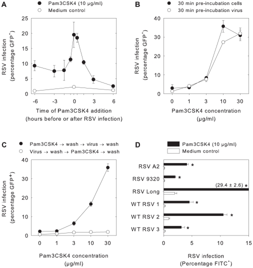
Figure 2. Pam3CSK4 mediates rapid and dose-dependent enhancement of RSV infection in B-LCL.
(A) B-LCL were incubated at 37°C with Pam3CSK4 (10 µg/ml) at 6 h, 3 h, 1.5 h, 0.5 h or 0 h before or 0.5 h, 1.5 h, 3 h, and 6 h after rgRSV infection. Pam3CSK4 treatment resulted in significantly increased rgRSV infection percentages in B-LCL at all time points (*p<0.005 with Student's t-tests with Bonferroni correction for multiple comparisons). (B) B-LCL or virus were pre-incubated with different concentrations of Pam3CSK4 for 30 minutes at 37°C before infection with rgRSV. Pam3CSK4 treatment resulted in significantly increased rgRSV infection percentages (p<0.05 for Pam3CSK4 concentrations ≥3 µg/ml), but no biologically relevant differences were detected between pre-incubation of cells or virus. (C) Cells were treated with different concentrations of Pam3CSK4 either directly before or after rgRSV infection. In all conditions free lipopeptide or free virus was washed away before proceeding to the next step. Pam3CSK4 treatment resulted in significantly higher percentages of infected cells (p<0.01, significant under Bonferroni correction for Pam3CSK4 concentrations ≥3 µg/ml). However, addition of Pam3CSK4 after rgRSV infection and subsequent removal of free virus had no effect. (D) B-LCL were incubated for 30 minutes at 37°C with or without Pam3CSK4 (10 µg/ml) before infection with different RSV strains. Pam3CSK4 treatment resulted in increased RSV infection percentages (p<0.05) for all strains tested. In all panels data are presented as percentages RSV-infected cells (means ± SD of triplicates) 20–24 hours after infection as determined by flow cytometry, using GFP expression in panels A–C and staining with FITC-labeled RSV-specific antibodies in panel D. Representative experiments out of three are shown.

