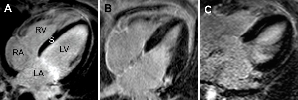
Figure 3.
LGE imaging in a normal subject who showed no signs of late enhancement in a four-chamber view (A). Patient suffering from ELD. Patchy pattern of delayed contrast enhancement (B and C). LA = left atrium, RA = right atrium, LV = left ventricle, RV = right ventricle, S = interventricular septum

