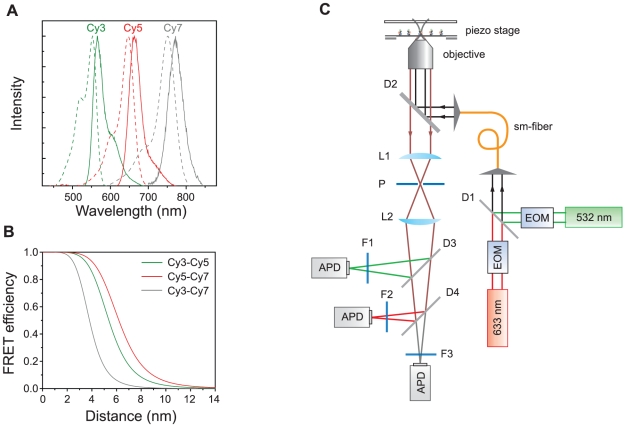
Figure 1. Dye selection and a confocal setup.
(a) Normalized emission (solid lines) and absorption (dash lines) spectra of Cy3 (green), Cy5 (red) and Cy7 (gray). (b) FRET efficiencies of the three FRET pairs as a function of inter-dye distances. R0 values of Cy3-Cy5 (green), Cy5-Cy7 (red) and Cy3-Cy7 (gray) pairs were calculated as 5.4-nm, 6.2-nm, and 3.8-nm, respectively. (c) A schematic diagram of ALEX three-color confocal setup. The setup was built based on an inverted microscope (TE2000-U, Nikon, Tokyo, Japan) equipped with a three dimensional piezo-stage (LP-100, MadCityLabs, Madison, WI). Two excitation lasers, a diode-pump solid state laser (532-nm, Excelsior-CDRH, Spectra-Physics, Santa Clara, CA) and HeNe laser (633-nm, HRP050, Thorlabs, Newton, NJ), were alternatively switched on and off by using electro-optic modulators (EOM, 350-50, Conoptics, Danbury, CT). To make sure that the two excitation lasers excite the same molecule, they were coupled into a single-mode fiber (460HP, Thorlabs). An oil-immersion objective (UPLSAPO 100×, Olympus, Tokyo, Japan) was used for both the excitation of molecules and the collection of fluorescence signals. The fluorescence signals are measured by using avalanche photo diodes (APD, SPCM-AQRH-14, Perkin Elmer, Wellesley, MA). The identities of other optics are: D1, a dichroic mirror (z532bcm, Chroma, Rockingham, VT); D2, dichroic mirror (z532/633rpc, Chroma); P, pinhole (P75S, Thorlabs); L1 and L2, lens (LAO-90.0-25.0/078, CVI, Irvine, CA); D3, dichroic mirror (640dcxr, Chroma); D4, dichroic mirror (740dcxr, Chroma); F1, bandpass filter (HQ580/60m-2p, Chroma); F2, bandpass filter (HQ680/60m-2p, Chroma); F3, bandpass filter (HQ790/80m, Chroma).

