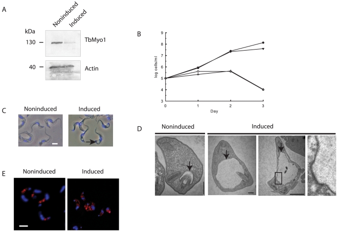
Figure 6. The effect of knock down of TbMyo1 in bloodstream forms of T. brucei.
Panel A. Western Blot analysis of TbMyo1 expression in a knock down cell line cultured for 24 h in the absence (noninduced) or presence of tetracycline (induced). Decreased expression of the TbMyo1 130 kDa protein is clearly visible in the induced cells. Each lane contained 5×10⁶ cells and a loading control was performed with antibodies against trypanosome actin. The loss of TbMyo1 had no effect on expression of actin. Panel B. The effect of knock down of TbMyo1 on the growth of bloodstream form RNAi cells. Two different bloodstream RNAi clones were grown in the presence (○, ▵) or absence of tetracycline (•, ▾). Panel C. The effect of the knock down of TbMyo1 on cellular morphology. The cells were cultured in absence (noninduced) or presence (induced) of tetracycline for 24 h, fixed and examined by phase contrast microscopy. A large phase-light vesicle or vacuole (arrow) located between the kinetoplast and nucleus (blue) was visible in the induced cells. Bar = 5 ųm. Panel D. Ultrastructural analysis of the effect of knock down of TbMyo1. The TbMyo1 RNAi bloodstream cells were cultured in the absence (noninduced) or presence (induced) of tetracycline for 24 h. The cells were processed for electron microscopy as described in the methods section. Sections through the flagellar pocket of a noninduced control cell and induced cells are shown. Enlargement of the flagellar pocket (arrow) is clearly visible in the induced cells. A noticeable feature of the enlarged pocket in TbMyo1 RNAi cells was the presence of the inward deformations of the flagellar pocket membrane but an absence of vesicles forming from the pocket. One of these regions (boxed) is shown at higher magnification. Bar = 500 nm. Panel E. Knock down of TbMyo1 affects the distribution of clathrin in cells. Bloodstream TbMyo1 RNAi cells were cultured in the absence (noninduced) or presence (induced) of tetracycline for 24 h. The cells were fixed and immunolocalizations were performed using an anti-clathrin antibody (red). The kinetoplast and nucleus are also shown (blue). Bar = 5 ųm.

