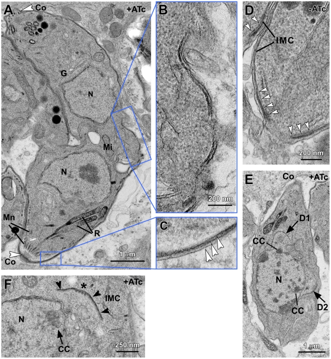
Figure 3. MORN1-KO mutants display a multi-headed, division phenotype.
(A–F) DNA content was assessed by flow cytometry with or without ATc for time periods as indicated. Defined populations representing 1N, 1.8N, 2.7N and 3.5N nuclear contents could be differentiated (B–D) and some are quantified in (E). Extracellular parasites lysed out after 48 hrs harbor large populations of 1.8N, and 3.5N nuclear content. (G) Extracellular parasites lysed out after 48 hrs were examined by immunofluorescence and identified multi-headed parasites with multiple nuclei (DAPI), mature subpellicular microtubules (α-tubulin) and IMC filament cytoskeleton (α-IMC3). (H) Single fluorescence channels and phase-contrast image of panel (G). (I) Extracellular parasites lysed out after 48 hrs were examined by scanning electron microscopy which identified basally conjoined parasites with 2, 4, 8 or 16 apical ends as indicated.

