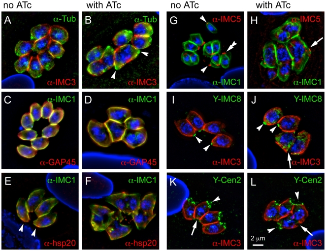
Figure 5. MORN1-KO parasites are defective in basal complex assembly.
MORN1-KO parasites were grown for 24 hrs in the absence (A,C,E,G,I,K) or presence (B,D,F,H,J,L) of ATc and subjected to immunofluorescence using either IMC1 or IMC3 antiserum, to highlight the IMC, in combination with the following antibody markers: (A,B) α-tubulin antibody, to highlight the subpellicular microtubular cytoskeleton; (C,D) GAP45 as a marker for maturation of the pellicle; (E,F) hsp20 as an independent IMC marker for pellicle maturation; (G,H) IMC5 antibody and (I,J) DD-YFP-IMC8 (Y-IMC8) as markers for the basal complex; (K,L) DD-YFP-Centrin2 (Y-Cen2) as a marker for the apical end and the basal complex. In all cases nuclear material was stained with DAPI. 1 µM Shield1 was added for 24 hrs to stabilize the DD domain fusion proteins. DD-tags were stained with α-FKBP12 (green). (A) Arrowheads indicate the two conoids present in the single cytoplasmic mass of a double-headed parasite. (E) Arrowheads indicate the basal accumulation of hsp20 in some uninduced parasites. (G-J) Arrowheads indicate the basal complex in some mother parasites, the double arrowhead indicates the contracting basal complex in a budding daughter, arrow indicates where the basal complex should have been assembled. (K,L) Arrowheads indicate the conoid at the apical end of some parasites whereas arrows indicate the basal complex (left panel), or where the basal complex should have been assembled (right panel). Single channel fluorescence panels are available in Supplementary Figure S3.

