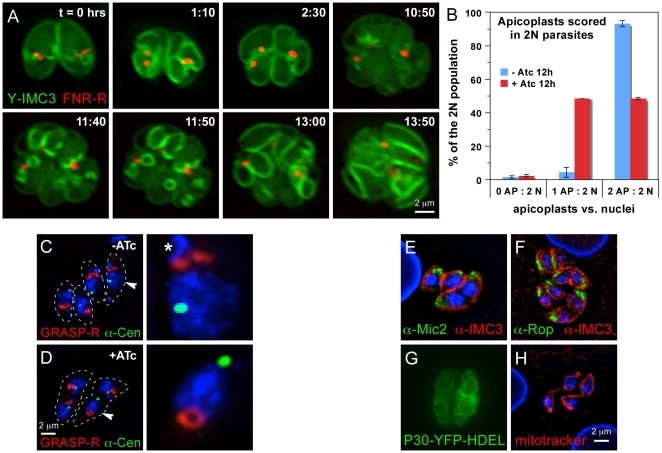
Figure 6. MORN1-KO parasites display a defect in apicoplast segregation and Golgi apparatus development.
(A) Selected panels of a time-lapse movie S1 of apicoplast division and parasite development in presence of ATc. ATc was added 12 hrs before t = 0 hrs. MORN1-KO parasites are expressing YFP-IMC3 and FNR-RFP marking the cortical cytoskeleton and the apicoplast, respectively. (B) The incidence of plastid loss after 12 hrs of ATc induction quantified in parasites with two nuclei (2N). Three categories are discerned: no plastid (0 AP), 1 plastid (1 AP) or 2 plastids (2 AP). (C,D) MORN1-KO parasites expressing the Golgi apparatus marker GRASP55-RFP, co-stained with DAPI and centrin antiserum. The parasites are outlined by a dotted line. In the right panels the nucleus plus Golgi and centrosome are 400% enlarged of the nuclei marked with an arrowhead in the left panel. Asterisk marks the nuclear content of the apicoplast. (E–H) Markers for the micronemes (α-Mic2), rhoptries (α-Rop), the endoplasmic reticulum (P30-YFP-HDEL) or the mitochondria (mitotracker) in the induced MORN1-KO parasites indicate normal development and segregation of these organelles.

