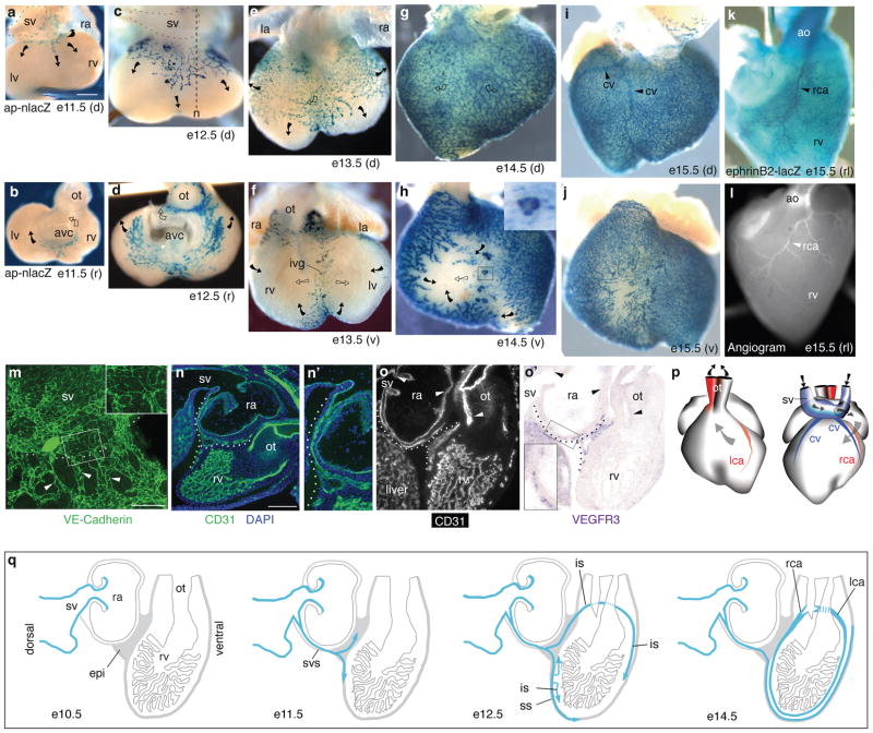
Figure 1. Coronary vessels sprout from the sinus venosus.
a–j. X-gal stained apelin-nlacZ hearts from embryonic days indicated, shown in dorsal (d), rostral (r), and ventral (v) views. Coronary vessels (blue nuclei) originate near the sinus venosus (sv, outlined in a,c) and spread dorsally (a, c, e, g) and around the atrioventricular canal (avc) (b, d) and outflow tract (ot) (d) to the ventral interventricular groove (ivg) (f). Arrows indicate direction of new growth on the surface (filled) or in deeper layers (open). Inset in h is close up of boxed blood island-like structure. la, left atrium; ra, right atrium; rv, right ventricle; lv, left ventricle. Scale bar (for a–l), 200 μm. k. X-gal-stained e15.5 ephrinB2-lacZ heart (right lateral (rl) view) showing right coronary artery (rca, arrowhead). ao, aorta. l. Angiogram of e15.5 heart showing right coronary artery (rca, arrowhead). m. Frontal section through e11.5 heart immunostained for VE-cadherin. Coronary sprouts (green, arrowheads) are continuous with the SV (above dashed line). Inset is close up of boxed region showing junction between SV and coronary sprouts. Scale bar, 100 μm. n. Sagittal section (approximate section plane shown in c) through e12.5 heart stained for CD31 (green) and with nuclear dye DAPI (blue). Coronary sprout (dotted line) extends from the sv and courses around right atrium (ra) to reach right ventricle (rv). n’. Close-up of coronary sprout in n. o,o’. Adjacent sections from e11.5 heart immunostained for CD31 (o) and probed for VEGFR3 mRNA (purple) (o’). Coronary vessels (dotted line) express VEGFR3, an angiogenic sprout marker, whereas vessels formed by vasculogenesis (sv, atria, outflow tract) do not (arrowheads). Inset in o’ is close-up of boxed region. Scale bar (for n,o,o’), 200 μm. p. Schematic of right (rca) and left (lca) coronary arteries and coronary veins (cv) at e15.5. Arrows show blood flow direction. q. Schematic sagittal sections showing coronary sprout (blue) progression during development. epi, epicardium; svs, sinus venosus sprouts; ss, superficial sprouts; is, invading sprouts.

