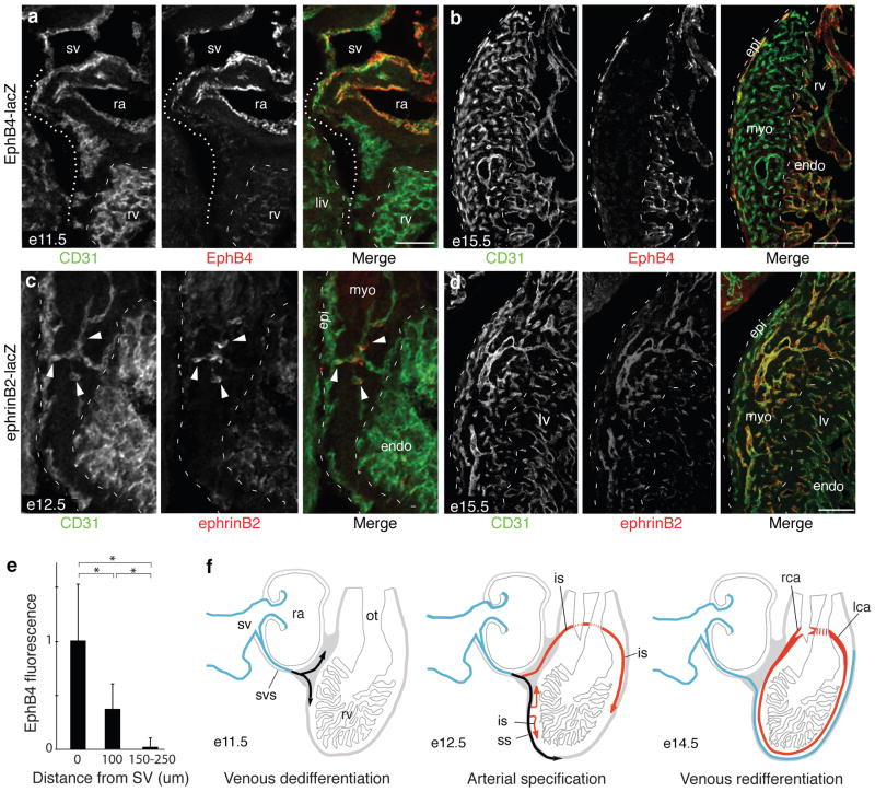
Figure 4. Downregulation of venous markers and induction of arterial markers during coronary artery development.
a. Sagittal section through heart of e11.5 EphB4-lacZ embryo immunostained for CD31 (green) and β-gal (EphB4, red). Coronary sprouts (dotted line) downregulate EphB4 as they migrate away from sinus venosus (sv). ra, right atria; rv, right ventricle; endo, endocardium; myo, myocardium; liv, liver. Scale bar, 100 μm. b. e15.5 EphB4-lacZ heart. Vessels on surface (epicardium, epi) have re-acquired EphB4 expression. Scale bar, 200 μm. c. Sagittal section through e12.5 ephrinB2-lacZ heart. Coronary sprouts that have invaded myocardium (green, arrowheads) upregulate artery/capillary marker ephrinB2 (red), whereas surface vessels (green, dashed line at left) do not. d. e15.5 ephrinB2-lacZ heart. EphrinB2 is expressed by all arteries and capillaries within myocardium but not by surface vessels. Scale bar, 100 μm. e. Quantification of EphB4 expression in coronary sprouts budding from sinus venosus (sv) at e11.5. Values are from double-stained hearts as in (a), normalized to CD31 staining at same position. Error bars, s.d. *, p<0.002 by Students t-test. f. Schematic showing changes in venous (blue) and arterial (red) marker expression during coronary development; black indicates dedifferentiated venous cells. svs, sinus venosus sprouts; ss, superficial sprouts; is, invading sprouts.

