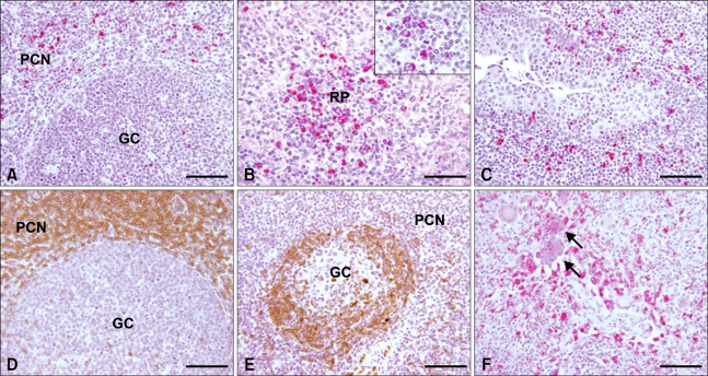
Fig. 1.
Immunohistochemical results showing expression of interukin (IL)-10 (A-C), CD3 (D), CD79a (E), and PCV2 antigens (F) in mandibular lymph nodes, spleen, and tonsil. (A) Mandibular lymph node; cytoplasmic staining of IL-10 antigens in marginal zone of lymphatic nodule. Scale bar = 140 µm. (B) Spleen; strong cytoplasmic staining of IL-10 antigen in splenic nodule. Scale bar = 56 µm. Inset: Higher magnification of IL-10 expression. (C) Tonsil; cytoplasmic staining of IL-10 antigen in and around tonsilar crypt. Scale bar = 56 µm. (D) Mandibular lymph node; strong immunohistochemical reaction to swine CD3, Scale bar = 140 µm. (E) Mandibular lymph node; strong immunohistochemical reaction to human CD79a. Scale bar = 140 µm. (F) Mandibular lymph node; presence of PCV2 antigen in the cytoplasm of macrophages and syncytial cells (arrows). Scale bar = 56 µm. GC: germinal center, PCN; paracortical nodule, RP: red pulp. (A), (B), (C) and (F) Alkaline phosphatase (red color) and hematoxylin stain. (D) and (E) Horse radish peroxidase (brown color) and hematoxylin stain.

