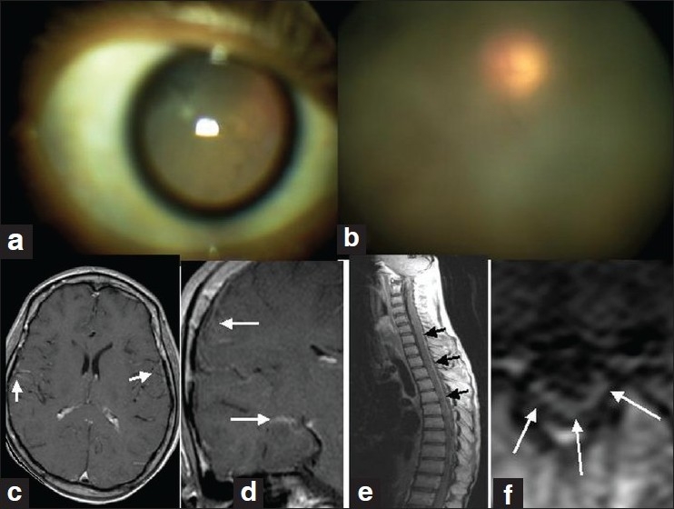
Figure 1.

a–e: (a) Dilated pupil; (b) hazy vitreous; (c and d) postcontrast axial and coronal T1W MRI of brain showing leptomeningeal enhancements (arrows); (e and f) post-contrast sagittal and axial T1W MRI of spine with extensive meningeal enhancement
