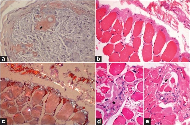
Figure 2.

Nerve and muscle biopsy. (a) Sural nerve biopsy showing part of one fascicle with large (*) and small amorphous endoneurial perivascular deposits of amyloid. (b–e) Muscle biopsy; abundant perimyseal and subperimyseal amyloid at the periphery of a fascicle, partly extending between the myofi bers (b); deposits are congophilic and display apple-green birefringence under the polarizer (c); amyloid deposits are also seen in the intramuscular nerve twig (*) (d) and in the muscle spindle (*) (E). (a and c): Congo red stain; c viewed under polarizer; b, d, and e: H and E stain; a–c: ×160; d and e: ×320, original magnifications)
