Figure 3.

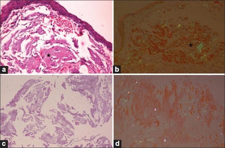
Conjunctival biopsy and vitreous material. (a) Conjunctival biopsy with large amyloid aggregates in the subepithelial tissue (*); (b) exhibiting typical congophilia and birefringence under the polarizer (*); (c) fluffy vitreous material; (d) displaying congophilia. (a and c: H and E stain; b and d: Congo red; a–d ×80 original magnification)
