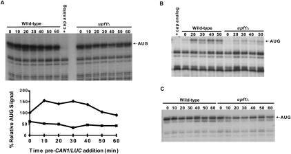
FIGURE 6.
Formation of 80S toeprints is diminished in upf1Δ extracts. (A) Cell-free translation extracts, prepared from WT and upf1Δ cells without MN treatment, were incubated at 18°C. CAN1/LUC mRNA (100 ng) was added at the indicated time points, and reactions were further incubated for 25 min, followed by addition of 2 mM cycloheximide (CHX) for 3 min. Reactions were frozen and subsequently analyzed by toeprinting. (Upper panel) The autoradiograph and (lower panel) the densitometry of the AUG band in WT (diamonds) and upf1Δ (squares) reactions normalized to the WT 0-min value. (B) Translation conditions were as in A, except that 2 mM CHX was added with 100 ng of CAN1/LUC mRNA at each time point, followed by incubation for 25 min. Lanes in A and B in which control reactions contained 2.7 mM cap analog are indicated. (C) Translation and incubation conditions were as in B, except that CHX was replaced with 2 mM GMP-PNP.

