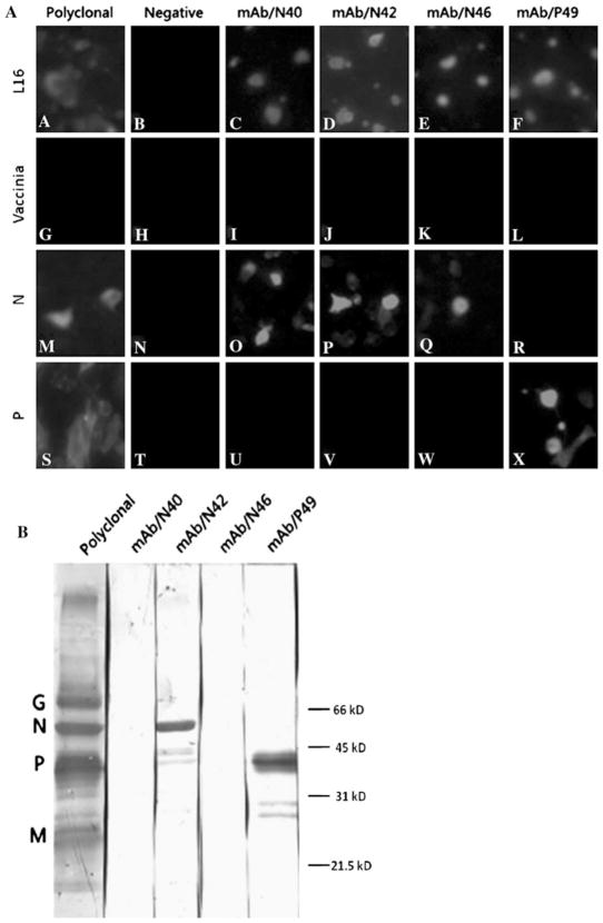
Fig. 1.
Characterization of RABV Mabs by immunofluorescence assay and by western blotting. In the immunofluorescent assay, a BSR cells were infected with RABV strain L16 or infected with vTF7-3 (MOI of 1), followed 1 h later by transfection with N- or P-expressing plasmid. At 48 h after infection or infection/transfection, cells were fixed with 80% acetone, reacted with individual Mabs, followed by goat anti-mouse IgG labeled with fluor 555. Panels A–F L16, cells infected with virus strain L16 only; Panels G–L Vaccinia, cells infected with vTF7-3 only; Panels M–R N, cells infected with vTF7-3 and transfected with N-expressing cDNA; Panels S–X P, cells infected with vTF7-3 and transfected with P-expressing cDNA. For Western blotting, b purified RABV L16 virions were subjected to SDS-PAGE, and the separated proteins were transferred to a nitrocellulose membrane. After blocking, the membrane was incubated with each of the Mabs, followed by incubation with peroxidase-conjugated secondary antibody. Reactivity of Mabs was determined by development with DAB. Each of the viral proteins recognized by the polyclonal antibodies is marked, and molecular weight markers were indicated as well

