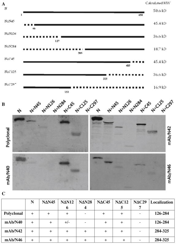
Fig. 2.
Epitope mapping of anti-N Mabs. RABV N with N-terminal or C-terminal deletions were constructed by PCR (a). The amplified fragments were cloned into pGEM-3Z at the PstI and XbaI sites as described previously [8, 26]. After in vitro transcription and translation, RABV N or mutant N proteins were used in immunoprecipitation reactions with individual anti-N Mabs. The precipitated proteins were subjected to SDS-PAGE and autoradiography (b). The results of the immunoprecipitation experiments are summarized in table form (c)

